
Cell-penetrating peptides (CPPs)—sometimes referred to as protein transduction domains (PTDs)—are short chains of amino acids (typically 5 to 30 residues) that can cross biological membranes.
Their defining feature is their ability to breach the cell’s protective lipid bilayer without permanently damaging the cell membrane or requiring specialized receptor machinery. Because of this, CPPs act as molecular “delivery vehicles,” ferrying attached molecular cargo directly into the cell’s interior.
How CPPs Work: Cellular Uptake
CPPs cross cell membranes primarily through two distinct mechanisms:
- Direct Translocation (Energy-Independent):At higher concentrations, certain peptides interact directly with the lipid bilayer, causing localized membrane destabilization, transient pore formation, or inverted micelle structures. This allows the peptide and its cargo to pass directly into the cytoplasm without using cellular energy ($ATP$).
- Endocytosis (Energy-Dependent):The cell actively engulfs the peptide (and its cargo) into a membrane-bound vesicle called an endosome. Once inside, the peptide must perform endosomal escape to enter the cytoplasm before the endosome fuses with lysosomes and degrades the payload.
Major Classes of CPPs
CPPs are typically categorized into three main chemical structural classes:
| Class | Key Characteristics | Typical Examples |
| Cationic | High density of positively charged residues (arginine and lysine). Electrostatic attraction binds them to negatively charged cell surface molecules. | Tat peptide (HIV-1 derived), Polyarginine ($R_8, R_9$) |
| Amphipathic | Distinct hydrophilic (polar) and hydrophobic (non-polar) regions, allowing dual interaction with aqueous fluid and lipid membrane cores. | Penetratin, Transportan |
| Hydrophobic | Primarily composed of non-polar amino acid residues that naturally integrate into lipid membranes. | Pep-1, K-FGF |
Key Biomedical Applications
- Targeted Drug Delivery: Coupling toxic therapeutic agents (such as chemotherapeutics) to targeted CPPs to improve intracellular concentration inside tumor cells.
- Gene Therapy & Nucleic Acid Delivery: Carrying large, fragile molecules like siRNA, mRNA, or CRISPR-Cas9 complexes across the lipid bilayer.
- Cell Imaging: Transporting contrast agents, quantum dots, or fluorescent dyes across membranes for high-resolution intracellular tracking.
What is Endosomal Escape?
When cell-penetrating peptides (CPPs) enter a cell via endocytosis, they become trapped inside membrane-bound vesicles called endosomes. For therapeutic cargo (such as mRNA, siRNA, proteins, or small-molecule drugs) to reach its site of action in the cytosol or nucleus, it must break out of these vesicles.
Endosomal escape refers to the step where CPPs permeabilize or rupture the endosomal lipid membrane, releasing their intact cargo into the cytoplasm before the endosome matures into a degrading lysosome.
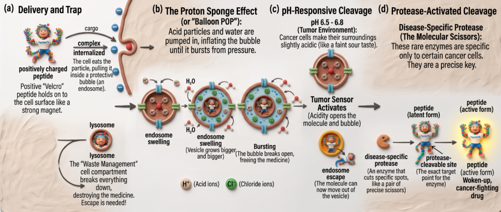
Notice in the diagram above how CPP-cargo complexes are enclosed within early endosomes. If endosomal escape does not occur, the endosome fuses with lysosomes where hydrolytic enzymes destroy the therapeutic payload.
Why Endosomal Escape is a Major Delivery Bottleneck
Despite high rates of cellular uptake, typically less than 1% to 5% of endocytosed CPP-cargo complexes successfully reach the cytosol in functional form. Endosomal entrapment represents the primary efficiency limit in non-viral intracellular delivery due to three factors:
- Lysosomal Degradation:Early endosomes rapidly mature into late endosomes, dropping their internal pH from ~6.5 to ~5.0. Late endosomes then fuse with lysosomes packed with proteases, nucleases, and lipases, rapidly destroying sensitive biological therapeutics.
- Physical Sequestration:The endosomal membrane forms a tight lipid barrier. Even if the cargo remains chemically intact, remaining physically locked inside an endosome prevents it from interacting with cytosolic machinery or translocating to the nucleus.
- Acid-Induced Inactivation:The accumulating acidity in maturing vesicles can denature or structurally destabilize delicate biological payloads (such as enzymes or antibodies) prior to escape.
Key Mechanisms of Endosomal Escape
Peptides exploit specific physical and chemical mechanisms to destabilize or escape endosomes:
1. The “Proton Sponge” Effect
- Mechanism: Peptides rich in weak bases (such as histidine residues with $pK_a \approx 6.0$) absorb incoming protons ($H^+$) pumped in during endosomal acidification.
- Outcome: To maintain electrical neutrality, chloride ions ($Cl^-$) flood into the vesicle alongside the protons, drawing water in via osmotic pressure. The endosome swells and eventually bursts, spilling its contents into the cytoplasm.
2. Membrane Disruption and Pore Formation
- Mechanism: As pH drops inside the endosome, amphipathic CPPs undergo structural changes that expose hydrophobic regions.
- Outcome: These hydrophobic domains insert into the endosomal membrane, forming transient toroidal or barrel-stave pores through which cargo diffuses into the cytosol.
3. Lipid Fusion and Phase Transition
- Mechanism: Positively charged CPPs interact electrostatically with anionic lipids (such as bis(monoacylglycero)phosphate, or BMP) found on internal endosomal membranes.
- Outcome: This interaction destabilizes normal lipid packing, inducing localized membrane fusion or inverted phase transitions that release the payload.
Strategies to Overcome the Bottleneck
To improve cytosolic delivery yields, drug delivery systems incorporate specific molecular modifications:
- Endosomolytic Sequences: Fusing CPPs with pH-sensitive viral peptides (e.g., HA2 from Influenza) that are inactive at neutral pH but adopt membrane-disrupting forms inside acidic endosomes.
- Histidinylation: Incorporating extra histidine residues into CPP backbones to increase buffering capacity and trigger osmotic swelling.
- Cleavable Linkers: Using pH-sensitive (e.g., hydrazone) or redox-sensitive (e.g., disulfide) chemical bonds between the peptide and cargo so the cargo separates cleanly once destabilization begins.